
Brain tumors


The origin of the word “tumour” is from the Latin “tumour” meaning “swelling”. In essence, a brain tumour is an overgrowth of the cells, which form the brain. This definition can also include tumors, which arise from cells of the brain’s surrounding structures like the meninges (brain linings) or the cranial nerves. Brain tumors can either be benign or malignant and are often not in themselves life-threatening. However, due to the limited space inside the bony skull (cranium), a benign or malignant tumour growing inside the skull can put pressure on healthy brain structures and cause damage.
Symptoms and diagnosis of a brain tumour
The type, size and location of the tumour dictate the kind of symptoms a patient may experience – these can include
- headache
- epileptic seizures (fits)
- visual disturbance
- speech disorder
- paralysis
- problems with movement
- memory disorders
The types of treatments available thus depend on the accurate specifications and symptoms of the tumor. In every case of a brain tumour it is of utmost importance that a diagnosis is made by a multidisciplinary team. That way, the best and most modern treatments can be chosen to increase each patient's benefit.
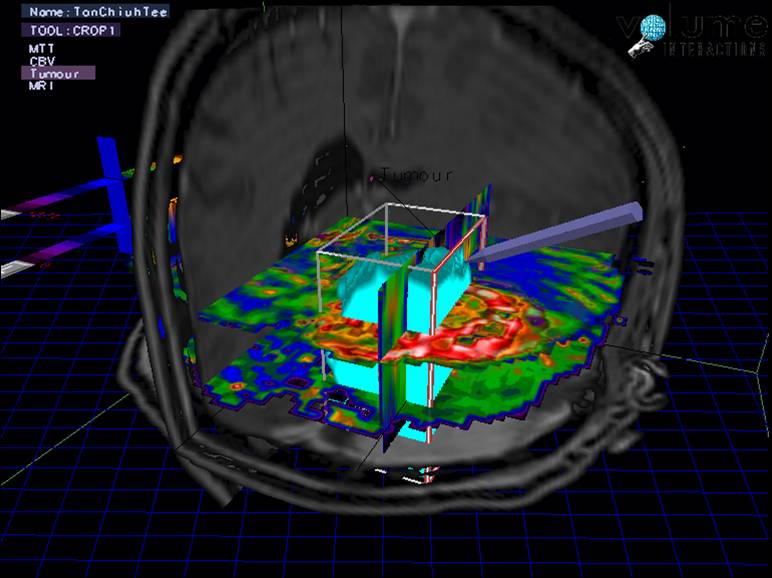
Diagnostics focuses on imaging with MRI (magnetic resonance imaging) and CT (computer tomography). In our clinic we have the most modern and powerful 3 T devices at our disposal, enabling imaging of high structural resolution and displaying functionally active regions (fMRI, functional MRI). This is of decisive importance for tumors in the vicinity of the speech region, the motor region or the visual center in order to be able to plan and perform an operation as precisely as possible.
In selected cases we perform at PET-CT or PET-MRI scan (Positron Emission Tomography) which may reveal how the tissue of a tumour is metabolically functioning. A PET scan uses a radioactive drug (tracer) to show the most active tumour regions as bright spots. This valuable information may then be used to focus the target of a microsurgical operation or the help in the decision making of other therapeutic steps.
Treatment of a brain tumour
In many cases, microsurgical resection is the best treatment for a brain tumour. The success of such an operation is measured in
- the degree of tumor resection set against
- the avoidance of damage to the surrounding healthy brain structures.
The risk of such a procedure can be successfully minimized through the control of two critical factors:
- choosing the right surgical approach to the target area and
- the use of appropriate operative techniques and equipment.
Procedure during surgery
The operating suites at the Hirslanden Klinik in Zurich are amongst the most advanced in the world. Cutting-edge intraoperative MRI (Magnetic Resonance Imaging) and CT (computed tomography) machines are used during tumour operations to control and optimize the resection.
High-precision navigation systems and intraoperative ultrasound are additionally available and are used to exactly confirm each phase of the operation in realtime 3D. In addition, a team of experienced neurologists and anesthesiologists monitor a range of brain functions during any operation in which functionally important parts of the brain are involved (electrophysiological monitoring), minimizing further the risk of neurological deficits after the operation.
In cases in which tumors involve the important language areas, we are able to perform awake craniotomies to permit optimal precision and preserve this critical function.

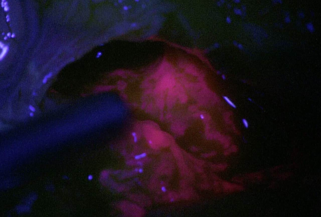
Use of fluorescent dyes during tumor removal
In some cases of malignant tumors, particuarly glioblastomas, recent research has shown that the precision of tumor resection can be significantly improved through the use of fluorescent dyes. The amino acid 5-ALA is given several hours before the operation and is taken up by tumor cells much more than in the surrounding, healthy cells. As a result, tumor tissue can be readily identified, glowing under the ultraviolet filter of the operating microscope.
With this technique, the all-important tumor margins can be easily visualized. We have found that a combination of intraoperative MRI and 5-ALA can further increase the precision of glioblastoma resection (Presentation at the Congress of Neurological Surgeons, San Francisco, Oktober 2014) and use this combination as standard with considerable success.
Use of endoscopes for tumor removal
In anatomically complicated areas such as the skull base or the ventricular system an endoscope offers unparalleled visual clarity. Essentially a tiny, moveable camera, the endoscope enables the inspection of difficult-to-access parts of the operating field and an unrestricted view of otherwise hidden pieces of residual tumor.
Combined with the use of special micro-instruments inserted via a tube shaft, this approach allows gentle and precise tissue dissection and maximal tumor resection. In the event that drilling is required in the region of the skull base, intraoperative CT can be used in combination with the navigation system with extreme precision to ensure safety zones around critical structures are not entered.
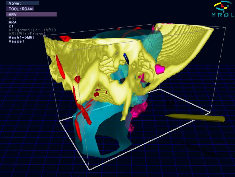
Three-dimensional computer simulation for surgery planning
We often use preoperative, 3D surgical planning with computer simulation of the individual stages of the operation, particularly in the region of the skull base. With this method we can safely define the exact spatial relationships of the important areas in each patient’s brain. We believe that the individual anatomy of a surgical target should be exactly understood before the surgery in order to chose a minimally invasive surgical strategy, avoiding exploratory tissue dissection und unnecessary tissue exposure. This is the key to a successful operation.
Preparation and follow-up treatment
Naturally, before and after each brain tumor operation you will be cared for by an interdisciplinary team of oncologists, radiation oncologists, neurologists, internal medicine specialists and other experts. Every patient with a brain tumor is discussed in a multidisciplinary team meeting (Tumorboard), a process which guarantees that the optimal treatment option is chosen – which may extend well beyond the tumor operation.